SKELETAL MUSCLE ORGANIZATION
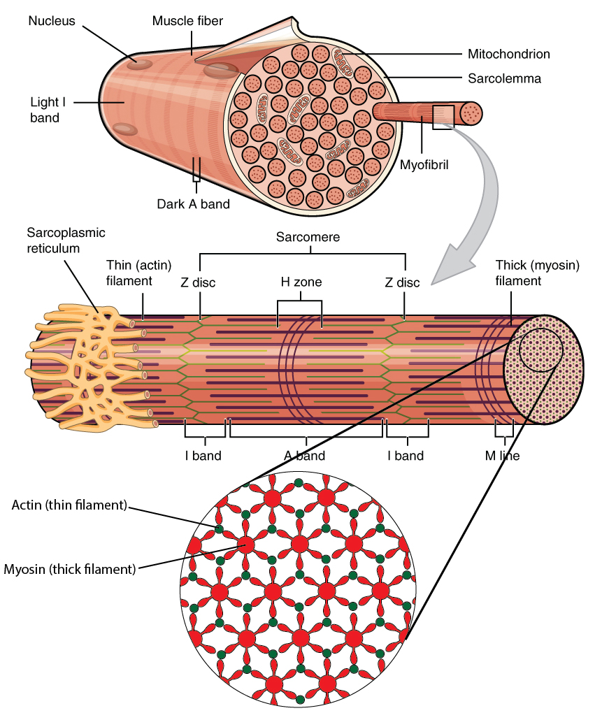
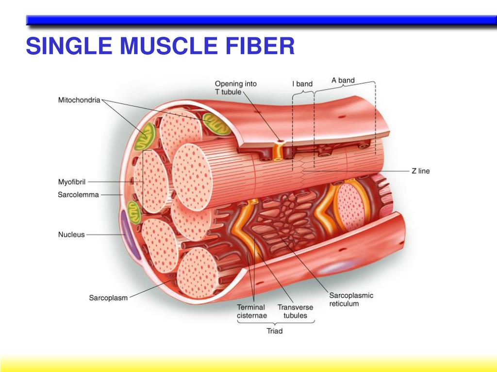
Muscle fibers are much longer than other cells as they were formed by many individual muscle cells fusing together when you were only an embryo. This makes the muscles strong, as any junctions between cells add a point of weakness.. Figure 3: A diagram of a section of a muscle fiber showing the intracellular structures of myofibrils, the.

Pin by Aline on Biology in 2022 Exercise physiology, Muscular system
Skeletal Muscle Fibers Because skeletal muscle cells are long and cylindrical, they are commonly referred to as muscle fibers (or myofibers). Skeletal muscle fibers can be quite large compared to other cells, with diameters up to 100 μ m and lengths up to 30 cm (11.8 in) in the Sartorius of the upper leg.

10.2 Skeletal Muscle Anatomy & Physiology
The fibers are relatively wide and very long, but unbranched. Fibers are formed from the fusion of thousands of precursor cells. This is why they are so long and why individual fibers are multinucleate (a single fiber has many nuclei). The nuclei are usually up against the edge of the fiber. There are striations in skeletal muscle. These are.
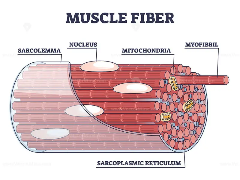
Muscle fiber structure and inner parts anatomical description outline
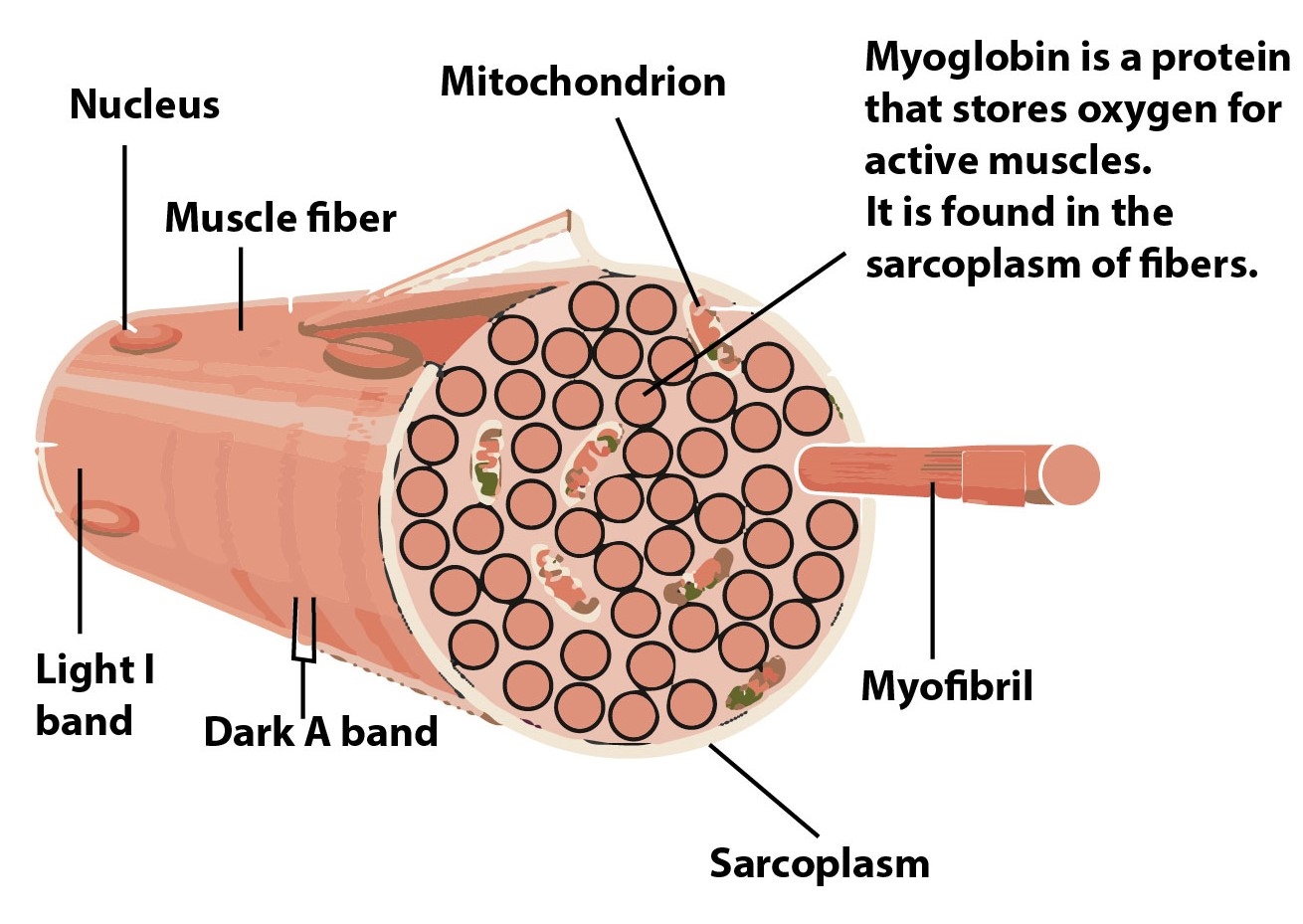
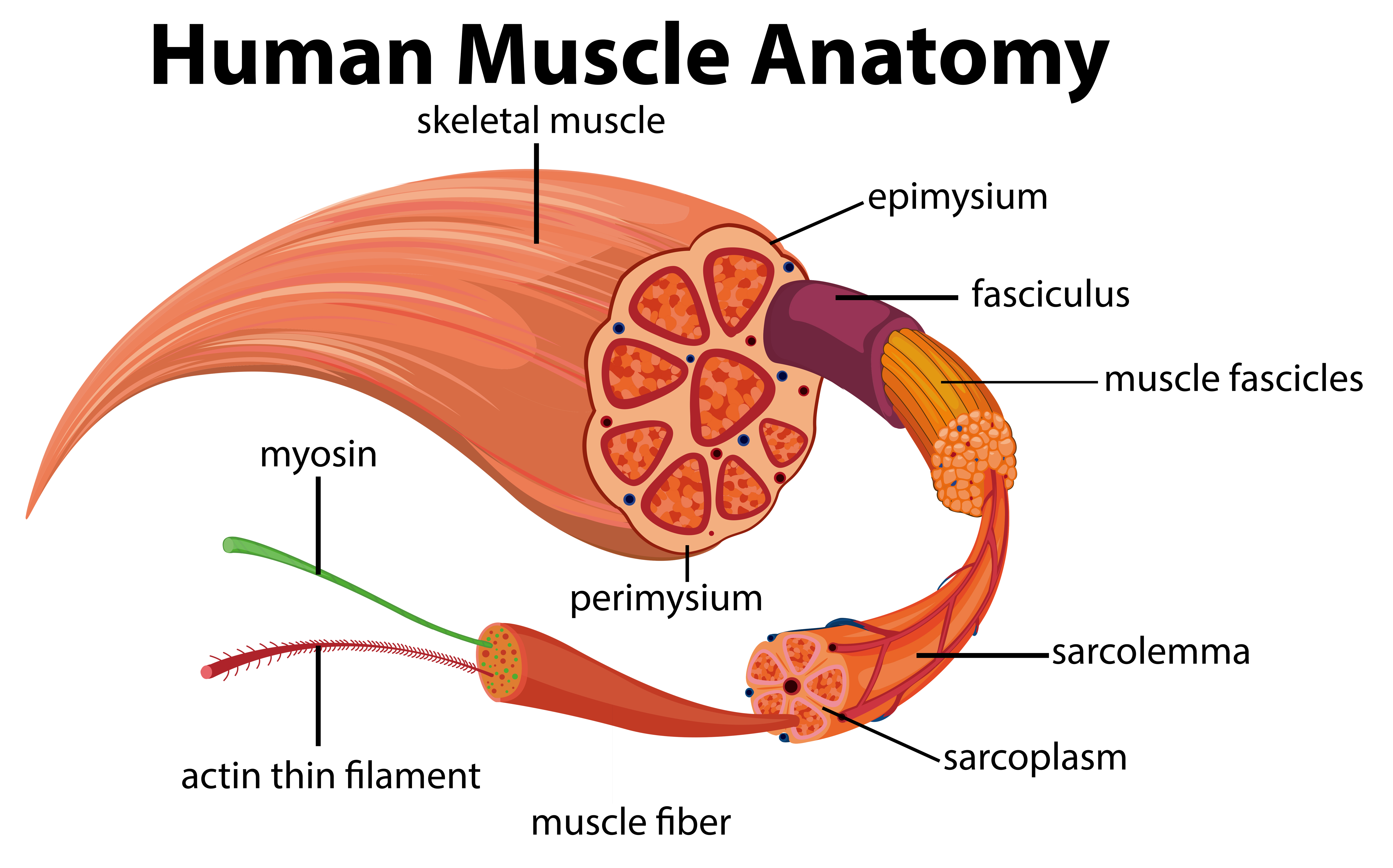
Muscles attach to bones directly or through tendons or aponeuroses. Skeletal muscles maintain posture, stabilize joints, support organs, control internal movement, and generate heat. Skeletal muscle fibers are long, multinucleated cells. The membrane of the cell is the sarcolemma; the cytoplasm of the cell is the sarcoplasm.

39 diagram of muscle fiber Trailer Wiring Diagram
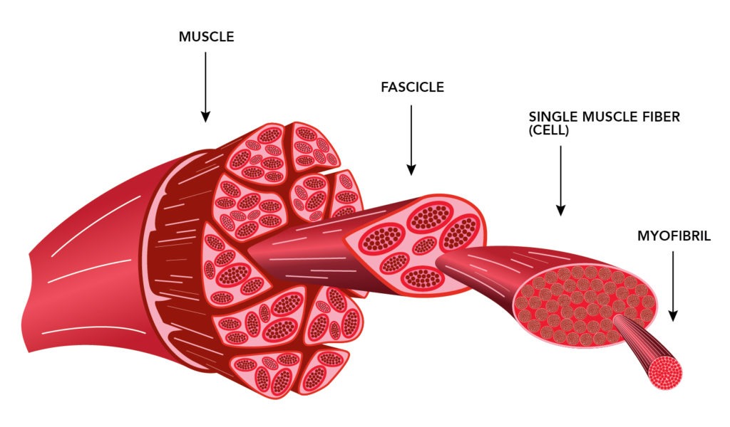
Each bundle contains multiple muscle fibres, which are formed when individual muscle cells fuse together. Muscle fibres contain tubular myofibrils that run the length of the fibre and are responsible for muscular contraction. The myofibrils can be divided into repeating sections called sarcomeres, each of which represent a single contractile unit.

1000+ images about A&P.2.Skin.Bone.Muscle on Pinterest Models, Muscle
Regardless of its morphology or type, muscle tissue is composed of specialized cells known as muscle cells or myocytes (myo- [muscle, Greek = mys]), commonly referred to as muscle fibers (all of these terms are interchangeable); this is due to their extensive length and appearance. Myocytes are characterized by protein filaments known as actin and myosin that slide past one another, producing.

SKELETAL MUSCLE PHYSIOLOGY STRUCTURE & TYPES OF MUSCLE FIBERS www
An oxygen debt is created as a result of muscle use. The three types of muscle fiber are slow oxidative (SO), fast oxidative (FO) and fast glycolytic (FG). SO fibers use aerobic metabolism to produce low power contractions over long periods and are slow to fatigue. FO fibers use aerobic metabolism to produce ATP but produce higher tension.
Muscle Fiber Diagram Unlabeled , Free Transparent Clipart ClipartKey
There are six actin molecules around a single myosin molecules and there are more than 100,000 sarcomeres (one myosin and six actin make 1 sarcomere) in a single bicep muscle fibre (a single cell) and 253000 such fibres in a young man's bicep. Now even if 10 percent of such fibres are stimulated at once there are more than 2530000000 sarcomeres.
Define the following structure Muscle, Muscle fibre, Myofibril
Relaxation of a Muscle Fiber Ca ++ ions are pumped back into the SR, which causes the tropomyosin to reshield the binding sites on the actin strands. A muscle may also stop contracting when it runs out of ATP and becomes fatigued. The release of calcium ions initiates muscle contractions. Watch this video to learn more about the role of calcium.
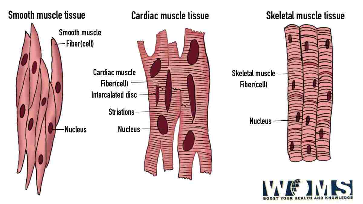
Diagram of Muscle Fiber 3 Types, Functions, and Anatomy WOMS
Identify areas of the skeletal muscle fibers Describe excitation-contraction coupling The best-known feature of skeletal muscle is its ability to contract and cause movement. Skeletal muscles act not only to produce movement but also to stop movement, such as resisting gravity to maintain posture.
How Heat Affects Muscle Fibers in Meat ThermoWorks
Fast-twitch, type II muscle fibers are further divided into type IIa and type IIb. Whereby type IIb (Fast-twitch glycolytic) fibers are more powerful but less resistant to fatigue than type IIa (Fast-twitch oxidative) fibers. For an image of muscle fibers from following paper: Muscle fiber type diversity revealed by anti‐myosin heavy chain.

9.3 Skeletal muscle fibers contain calciumregulated molecular motors
Diagram the process of cross-bridge cycling; The Neuromuscular Junction. The process of muscle contraction begins at the site where a motor neuron's terminal meets the muscle fiber—called the neuromuscular junction (NMJ). Every skeletal muscle fiber in every skeletal muscle is innervated by a motor neuron at a NMJ. Excitation signals from.
PPT MUSCLES AND HOW THEY MOVE PowerPoint Presentation, free download
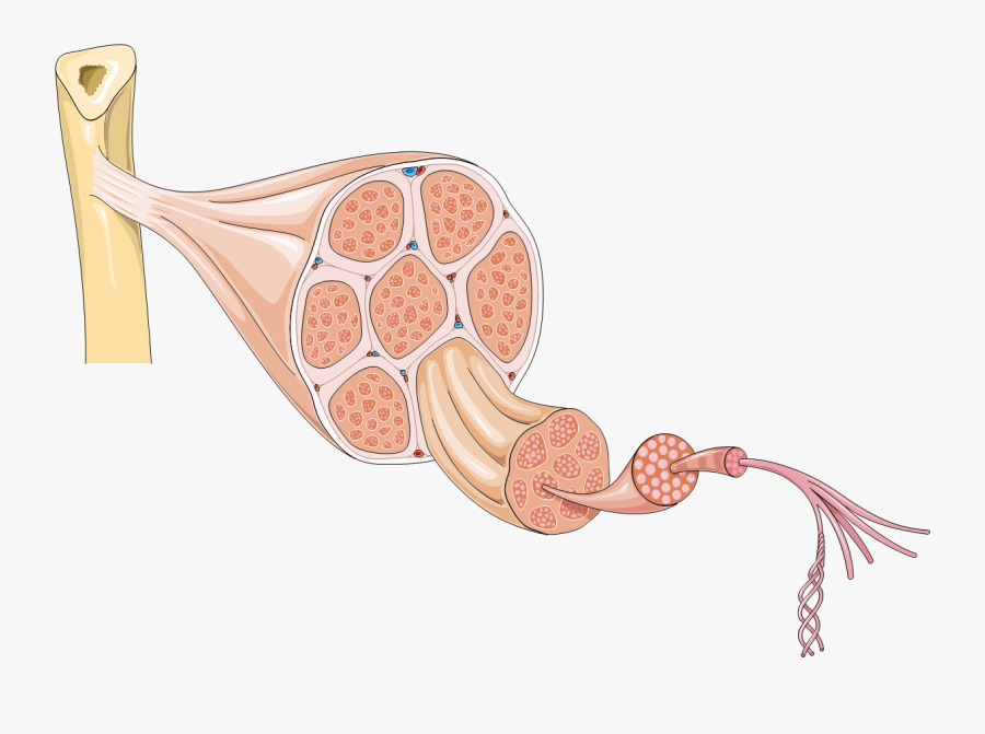
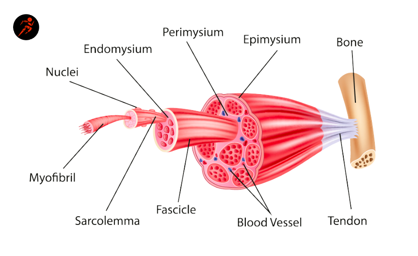
Figure 1. The Three Connective Tissue Layers. Bundles of muscle fibers, called fascicles, are covered by the perimysium. Muscle fibers are covered by the endomysium. Each skeletal muscle is an organ that consists of various integrated tissues. These tissues include the skeletal muscle fibers, blood vessels, nerve fibers, and connective tissue.
Muscle Fibers Explained Type I and Type II (Slow & Fast Twitch
Skeletal muscles (commonly referred to as muscles) are organs of the vertebrate muscular system and typically are attached by tendons to bones of a skeleton. [1] [2] The muscle cells of skeletal muscles are much longer than in the other types of muscle tissue, and are often known as muscle fibers. [3]
Muscle Fiber Vector Art, Icons, and Graphics for Free Download
Sliding filament model of muscle contraction Muscle contraction Neuromuscular junction and motor unit Osmosis Muscles high-yield notes offers clear overviews with striking illustrations, tables, and diagrams. Make learning more manageable.

Muscle Fiber Contraction and Relaxation Anatomy and Physiology
Skeletal muscle Each one of your skeletal muscles is made up of hundreds to thousands of muscle fibers that are tightly wrapped together by connective tissue. Each muscle fiber contains.